Optophysiology: interferometric imaging of neural signals and cell metabolism

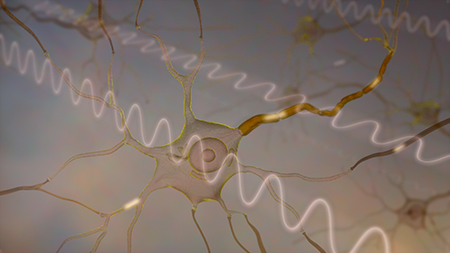
Neural signals involve rapid changes of the cell potential, which affect the cell membrane tension. As a result, cells slightly deform when trans-membrane voltage changes, which provides an intrinsic biomarker of the neural activity. We developed a wide-field interferometric imaging sensitive to such minute deformations, and we are working now on implementation of this approach to label-free all-optical monitoring of the neural signals in-vivo – in the retina and in other optically accessible tissues.
Sub-nanometer precision of the quantitative phase imaging opens the door to optophysiology - an optical alternative to traditional electrophysiology: non-invasive label-free optical detection of neural signals, as well as other metabolic changes in cells and tissues. We are working on two approaches to quantitative phase imaging: full-field interferometry in transmission, and phase-resolved optical coherence tomography in reflection.
We study the mechanisms of electro-optical coupling and develop biological applications of this technology, including mapping the neural activity in the retina during light exposure, optical thermometry, and others.

