Dave's Reoviridae Update 2008!
This site is intended for fellow virophiles! Hope you find it helpful!
Links to Previous Students' Reoviridae Webpages
Viral Morphology:
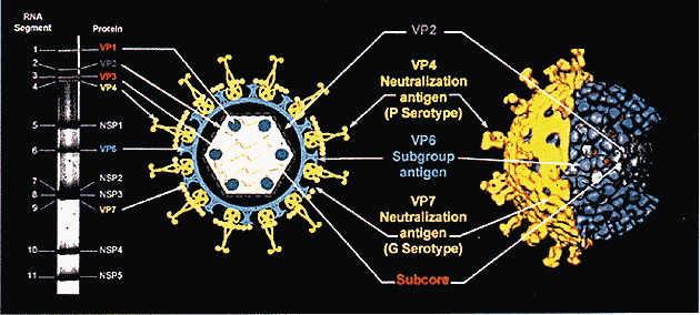
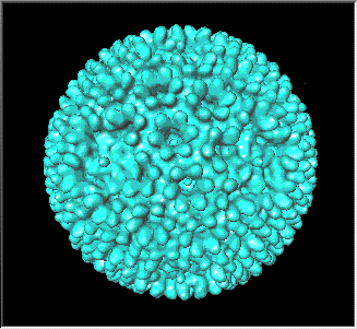
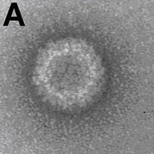
Rotaviruses are roughly 100 nm particles with three capsids, a feature not seen in any other human virus family (Knipe and Howley 1918 [Throughout this page, this citation refers to Fields Virology]). The outer two capsids, the outer comprised of VP4 and VP7 and the inner of VP6, have a triangulation number of T=13l, another unique aspect of reoviral morphology. In the outer two layers are channels connecting the innermost capsid (the inner core) to the “outer world” through which newly synthesized mRNA is extruded (Knipe and Howley 1924). The inner core houses all the enzymatic functions of the virus and has a T=1 morphology created by dimers of VP2, leading some to call it the unique T=2 (Knipe and Howley 1925).
Rotavirus capsid morphology(1)
Electron micrograph of rotavirus(2)
Genome Organization:
The rotaviral genome is made up of 11 segments of double-stranded RNA (dsRNA). Each segment contains one open reading frame (except for segment 11, which contains two [Knipe and Howley 1927]) sandwiched between 5’ and 3’ conserved sequences. The 3’ end of the genome encodes translation enhancers and is not poly adenylated (Knipe and Howley 1926). In the capsid, the segments are hydrogen bonded to one another end-to-end and the 5’ end of the positive sense strand has a 5’ cap. The logistics of packaging a segmented genome can be quite overwhelming, with each segment needing to encode the same sequence to be recognized by the polymerase in addition to a unique segment that will distinguish it from the other segments, a signal possibly encoded in the highly conserved noncoding regions. In addition to shift, these segments are also able to undergo rearrangement in which bases are added or deleted (Knipe and Howley 1927). However, the rearrangement usually leaves the open reading frames intact (Knipe and Howley 1928).
TABLE 53.4 Rotavirus proteins |
||||||||||||||||||||||||||||||||||||||||||||||||||||||||||||||||||||||||||||||||||||||||||||||||||||||||||||||||
|
||||||||||||||||||||||||||||||||||||||||||||||||||||||||||||||||||||||||||||||||||||||||||||||||||||||||||||||||
Adsorption:
Rotaviruses attach to target cells using VP4 or its cleavage product, VP5*, but the initial binding is very nonspecific, involving cellular sialic acid (Knipe and Howley 1929). The specificity of rotaviruses for differentiated enterocytes of the small intestine probably comes from the interaction of VP8*, another cleavage product of VP4, with 2-O-methyl α-SA (Knipe and Howley 1930).
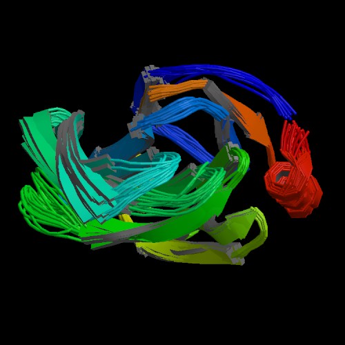
Rhesus rotavirus VP4(3)
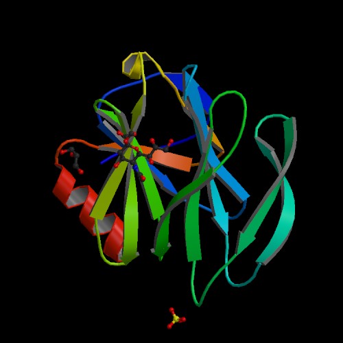
Rhesus Rotavirus VP4 complexed with 2-O-methyl alpha sialic acid(4)
Penetration and Uncoating:
Rotavirus entry into the cell involves a series of conformational changes in the capsid proteins after binding with cellular receptors such as integrins. The detailed mechanism of penetration, however, remains unclear. It appears, though, that it involves Ca2+-dependent fusion with the lysosome membrane (Knipe and Howley 1931).
Transcription:
Transcription of the rotavirus genome is accomplished by an endogenous RNA-dependent RNA polymerase made of a complex of VP1 and VP3 found at each of the 5-fold axes of the inner core. In the triple layered particles, which have all three capsids, the transcriptase complex is inactivated, but removal of the outer capsid activates this complex. Transcription proceeds using the negative strand as a template to make positive sense RNA transcripts (capped but not poly adenylated), which are released through the channels in the capsid (Knipe and Howley 1932).
Translation:
VP4 and VP7 are both assembled on the rough endoplasmic reticulum, whereas the other viral proteins are synthesized on free ribosomes (Knipe and Howley 1932-3).
Replication:
Creation of dsRNA occurs in aggregations of subviral particles in inclusion bodies called viroplasms. Positive sense single-stranded RNA is the template for synthesis of its negative sense complement. During the entire process, the newly synthesized dsRNA remains within the partially uncoated virions in which they were produced. Interestingly, it appears that there are separate transcripts that are used for replication and translation (Knipe and Howley 1933).
Encapsidation:
Although the mechanism by which encapsidation of each—and only one of each—segment occurs is unclear, several hypotheses have been advanced. The first holds that polymerase/replication intermediates serve as nucleation sites for the formation of the inner core. A second model maintains that mRNAs are inserted into ready-made cores. The last one claims that VP1, VP3, and two copies of VP2 form tetramers, each responsible for transcribing a specific segment of the genome. After transcription of the segments, interactions between the nascent nucleic acids drives assembly of the icosahedron (Knipe and Howley 1935).
Virion Assembly:
Although subviral particles are formed in viroplasms, they end up budding through the endoplasmic reticulum, transiently giving maturing particles an envelope, a step not seen in any other viral family let alone any other genus of Reoviridae (Knipe and Howley 1935). Eventually, the envelope is replaced by the outermost capsid. One important viral protein in the assembly process is NSP4, which is a membrane bound protein assembled on the endoplasmic reticulum facilitating budding of the double-layered particles its lumen. In addition, this protein is thought to be involved in removal of the transient envelope by upregulating the intracellular concentration of calcium ions (Knipe and Howley 1936-7).
Egress:
Assembled virions leave cells by permeablizing the cellular membrane, resulting in cell lysis (Knipe and Howley 1938).
Diagnosis:
Because the syndrome induced by rotavirus is not unique, many causes are on the differential diagnosis of gastroenteritis. Therefore, diagnosis is not syndromic, but relies on either virus/viral antigen detection or a serologic response. Many methods have been used in the detection of rotavirus virions and viral proteins, including electron microscopy, ELISA, RT-PCR, viral culture, and flow cytometry (Knipe and Howley 1950). To detect serologic response, complement fixation, immunofluorescence, ELISA, and other methods have been used. It should be noted that detection of rotavirus or an immune response to rotavirus does not confirm an etiological link between the patient’s gastroenteritis and the virus since there are many inapparent infections (Knipe and Howley 1951).
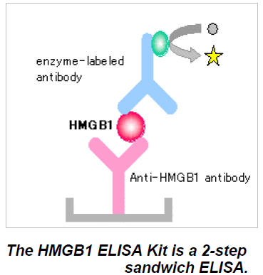
Example of enzyme-linked immunosorbent assay scheme used in diagnosis (ELISA)(5)
Treatment:
As rotavirus can cause severe dehydration due to gastroenteritis, the goal of all treatment is to replace lost fluids, that is to say treatment is symptomatic. There are no antivirals for rotavirus infection. Since much of the mortality due to rotavirus (between 500,000 and 1,000,000 deaths per year in children under 5 [Knipe and Howley 1917]) occurs in regions of the world with limited health infrastructure, it is important to find low-technology, efficacious solutions to diarrhea. In the late 1960’s, researchers discovered that the intestine has an additional way of absorbing sodium, namely sodium/glucose cotransports, which bring in one molecule of sodium for every molecule of glucose absorbed. Because these proteins are not affected by rotavirus, supplying water with glucose as well as sodium reestablishes the osmotic pressure across the gut. Later research has also discovered similar sodium cotransports that use amino acids as well, leading the WHO to include both glucose and amino acids in their oral rehydration therapy (ORT) solutions(6). One factor that has limited the impact this has had on people has been the availability of clean water. For many of the people for whom rotavirus represents a life-threatening disease, clean water can be hard to come by making it hard to avoid reinfection with diarrhea-causing pathogens.
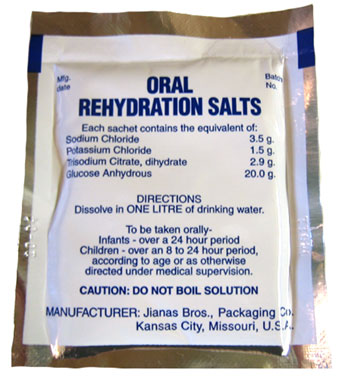
Oral Rehydration Therapy (ORT)(7)
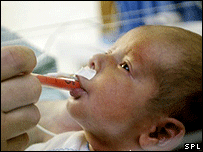
Dehydrated child receiving ORT(8)
Prevention:
RotaShield, the original vaccine for rotavirus, was a tetravalent reassortant rhesus rotavirus vaccine. After nearly a year in production, it was pulled from the American market when links were drawn between it and intussusception, a condition in which the intestine telescopes in on itself, killing the gut and possibly leading to lethal bacterial infection(9). This decision by the American Advisory Committee on Immunization Practices to withdraw its endorsement of the vaccine had large consequences for those suffering from rotavirus diarrhea in developing countries. Not only did the loss of the United States as a market drastically undermine the producer’s ability to make a profit, but also it led to public relations maneuvering within the developing countries to stop supplying the vaccine. After all, what government would want to supply to its people a vaccine deemed too dangerous for the American public? What such logic did not take into account was the differing levels of risk due to the vaccine in the two settings. In a country such as the United States, in which only 20 to 40 people die per year from rotavirus (Knipe and Howley 1917), the threat of death due to intussusception—however small—was unacceptable. In developing countries around the world, though, the calculus definitely was in favor of continued use of the vaccine (51 deaths due to intussusception/yr vs 500 deaths due to rotavirus gastroenteritis/yr). Adding to the controversy is the fact that reexamination of the data reveals that the link between intussusception and rotavirus vaccination may not be near as strong as once thought. In 2006, a new, monovalent vaccine called RotaRix was licensed in a number of countries (the US not included), ending the seven year drought of an effective rotavirus vaccine (Knipe and Howley 1956-7). Soon after that, RotaTeq, a human-bovine reassortant, pentavalent, live attenuated, oral vaccine, was approved for use in the United States(10)(11).
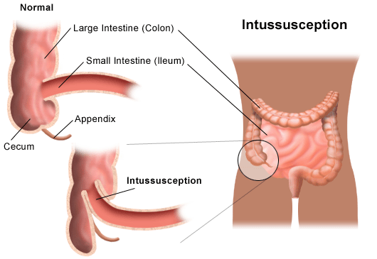
Intussusception(12)

RotaTeq(13)

RotaRix(14)
Immunity:
Evidence on the relative importance of local and systemic immunity has been very conflicted, with some studies holding that local intestinal immunity is more important than systemic and others maintaining that serum antibody is a good indicator of resistance to infection (Knipe and Howley 1946). However, there does seem to be some protective effect of rotavirus antibodies against any rotavirus infection and complete protection against serious infection, whatever the relative importance of local and systemic immunity may be (Knipe and Howley 1947).
Clinical Picture:
Rotavirus infection can range from subclinical to life-threatening dehydrating disease (Knipe and Howley 1948). Gastroenteritis due to rotavirus infection can involve both diarrhea, lasting around 5 days, and vomiting, lasting roughly 2.5 days. It is interesting to note that one of the rare side effects of infection with wild type rotavirus is intussusception, raising concerns that the intussusception seen with the first rotavirus vaccine may not have been due to the specific strain used, but merely infection with any rotavirus (Knipe and Howley 1949).

"Diarrhea is very dangerous--Give your child oral rehydration solution"(15)
Pathogenesis:
Rotavirus replicates near the tips of villous enterocytes and can result in everything from no histopathological change to villus blunting and crypt hyperplasia. Interestingly, there is often no correlation between severity of disease and degree of pathologic changes and many times diarrhea onset occurs before pathological changes in the gut. These two observations made clear that there must be additional mechanisms to induce diarrhea instead of or in addition to destruction of the architecture of the small intestine. Many of the rotavirus proteins have been linked to rotavirus’s pathogenesis in animal models, among them NSP4, the first viral enterotoxin discovered (Knipe and Howley 1940). In addition, rotavirus stimulates the enteric nervous system, another way in which the virus is thought to result in diarrhea (Knipe and Howley 1941).
Epidemiology:
Although rotavirus is ubiquitous, the impact it has on people is largely determined by their ability to nourish and rehydrate themselves long enough for their immune system to clear the infection from their body. In resource-poor settings, people might not have access to water and food to replace what is lost from diarrhea and vomiting. In the US, for example, the virus is estimated to cause between 20 and 40 deaths per year, whereas its worldwide under 5 mortality between 500,000 and 1,000,000 deaths per year (Knipe and Howley 1917). However, because it can cause serious diarrhea anywhere, its costs are significant--$400 million in direct medical costs and $1 billion in societal costs in the US every year (Knipe and Howley 1941). Although rotavirus can infect adults, clinically apparent Group A rotavirus disease is rare because of lingering antibodies to the virus. Infection with Group B rotavirus, though, has been linked to epidemics of severe adult gastroenteritis in China. Rotavirus is transmitted fecal-orally and possibly by a respiratory route, as well. It is important to note that many rotavirus infections are acquired nosocomially, a route of transmission made much easier by the virus’s stability in the environment. It appears that fomites are the major normal way in which rotavirus is transmitted, as opposed to more typically waterborne pathogens such as E. coli (Knipe and Howley 1943). In temperate climates, patterns of rotavirus infection show a seasonality, peaking in the colder months. However, in more tropical regions, less seasonality is observed (Knipe and Howley 1944). Not only does malnutrition predispose people to rotavirus infection, but it can also aggravate malnutrition by interfering with absorption of macromolecules across the intestine.
Map of Mortality burden of rotavirus (each dot=5000 deaths)(16)
Taxonomy:
The family Reoviridae is comprised of twelve genera, four of which contain human pathogens—Orthoreovirus, Rotavirus, Coltivirus, and Orbivirus (Knipe and Howley 1854). Members of the genus orthoreovirus, which contains pathogens of mammals, birds, and reptiles, consist of 10 segments of dsRNA and are divided into fusogenic and nonfusogenic viruses based on their ability to induce syncytia, an unusual property for naked viruses (Knipe and Howley 1855). This genus, and the family as a whole, get their name from the acronym of respiratory, enteric, and orphan; the first reoviruses isolated were found in human respiratory and enteric tracts but were not associated with disease, and were hence called orphan. To this day, only one human orthoreovirus has been isolated and it is not associated with disease (Knipe and Howley 1856). The genus orbivirus consists of 10 segmented bird and mammal arthropod borne viruses (Knipe and Howley 1976). Although there are a handful of zoonoses, the most important orbiviruses are bluetongue virus, which causes lesions and a blue tongue in domestic animals, and African horse sickness virus, which is a lethal disease of horses (Knipe and Howley 1975). Coltiviruses have twelve segments of dsRNA and are tickborne viruses causing meningitis, encephalitis, and other neurological symptoms(17). Rotaviruses, as discussed in detail in the rest of this site, have 11 segments of dsRNA and are fecal-orally transmitted, leading to gastroenteritis in both mammals and birds (Knipe and Howley 1924).
Colorado tick fever virus(18)
Eyach virus(19)
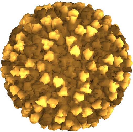
Bluetongue virus (an orbivirus of domesticated animals)(20)
Changuinola (Knipe and Howley 1977)Rotavirus A (Knipe and Howley 1919)
Rotavirus B (Knipe and Howley 1919)
Rotavirus C (Knipe and Howley 1919)
Orthoreovirus(21)
Human Reovirus(22)
Colorado Tick Fever:
Colorado tick fever, also called mountain fever or American mountain fever, is a member of the genus Coltivirus and is transmitted by the tick Dermacentor andersoni, which lives in the Rocky Mountains. Incidence is highest in May and June, but infection can occur any time between March and September. Usually self-limiting, after 3 to 6 days, patients develop a sudden fever, accompanied by severe myalgia, arthralgia, headache, nausea, and vomiting. Confirmatory tests include complement fixation and immunofluorescence. Treatment entails removing the tick and treating symptomatically any symptoms that occur. Possible complications include meningitis, encephalitis, and hemorrhagic fever, although these are very rare. Prevention of infection involves taking proper precautions, such as tucking long pants into socks and removing ticks from skin and clothing as soon as they are discovered(23).
Colorado Tick Fever Virus(24)
1) Reoviridae is the "multiplicity family" because it has multiple capsids and multiple segments.
2) Because the genome is comprised of dsRNA, the virus needs to find a way to sequester its genome so it does not provoke an interferon response. It accomplishes this by never fully uncoating the virion, but instead synthesizing the mRNA within the capsid (Knipe and Howley 1863).
3) Rotavirus is the most important cause of severe diarrhea worldwide.
4) Orthoreovirus vectors have been suggested in the treatment of cancer because the activated ras pathway makes cancer cells susceptible to lysis by orthoreoviruses (Knipe and Howley 1899).
5) Rotavirus is transmitted fecal-orally.
6) Rotavirus is treated with oral rehydration therapy, which reestablishes the osmotic pressure across the gut, decreasing the amount of water lost to diarrhea.
7) Reoviridae gets its name from an acronym of the words respiratory, enteric, and orphan because the first reoviruses were found in the respiratory and enteric tracts but could not be linked with any human disease.
8) There are currently two vaccines for rotavirus. One is licensed in the United States (RotaTeq), while the other is marketed in many developing countries (RotaTeq).
9) Under the electron microscope, reoviruses look like Ritz crackers.
10) The family as a whole has quite the impressive host range, able to infect plants, insects, fungi, and animals (fish, molluscs, birds, reptiles, and mammals) (Knipe and Howley 1854).
Lucille Packard Children's Hospital Rotavirus Page
Clinical Trials Involving Rotavirus
ICTV Reoviridae Classification Page
Utah Department of Health Site on Colorado Tick Fever
Halasz, Peter, et al. "Rotavirus Replication in Intestinal Cells Differentially Regulates Integrin Expression by a Phosphatidylinositol 3-Kinase-Dependent Pathway, Resulting in Increased Cell Adhesion and Virus Yield."
The way in which cell surface integrins interact with the extracellular matrix largely determines the growth and survival of enterocytes. In this study, the authors noted changed integrin expression after rotavirus infection. They were able to determine that a2b1 and b2 are upregulated, while aVb3, aVb5, and a5b1 are downregulated. Further, the authors determined that integrin regulation is dependent on phosphatidylinositol 3-kinase (PI3K), which increased adherence of infected cells to collagen and increased virus production.
Halasz, Peter, et al. "Rotavirus Replication in Intestinal Cells Differentially Regulates Integrin Expression by a Phosphatidylinositol 3-Kinase-Dependent Pathway, Resulting in Increased Cell Adhesion and Virus Yield." Journal of Virology 82.1 (2008): 148-160. 9 Mar. 2008 <http://jvi.asm.org/cgi/content/full/82/1/148?view=long&pmid=17942548>.
Beau, Isabelle, et al. "A Protein Kinase a-Dependent Mechanism by Which Rotavirus Affects the Distribution and MRNA Level of the Functional Tight Junction-Associated Protein, Occludin, in Human Differentiated Intestinal Caco-2 Cells."
Properly functioning tight junctions (TJ), which form an impenetrable barrier between the apical and basal portion of enterocytes, are crucial in establishing the osmotic pressure that allows for water reabsorption across the gut. In this study, the authors found that one of the TJ proteins, occludin, was downregulated at the transcriptional level by rotavirus infection in rhesus monkeys.
Beau, Isabelle, et al. "A Protein Kinase a-Dependent Mechanism by Which Rotavirus Affects the Distribution and MRNA Level of the Functional Tight Junction-Associated Protein, Occludin, in Human Differentiated Intestinal Caco-2 Cells." Journal of Virology 81.16 (2007): 8579-8586. 9 Mar. 2008 <http://jvi.asm.org/cgi/content/full/81/16/8579>.
Beau, Isabelle, Arnaud Berger, and Alain L. Servin. "Rotavirus Impairs the Biosynthesis of Brush-Border-Associated Dipeptidyl Peptidase IV in Human Enterocyte-Like Caco-2/TC7 Cells."
In addition to their findings in the previous article, Beau’s lab also found that rhesus rotavirus infection downregulates translation of dipeptidyl peptidase IV (DPP IV), an important protein in the catabolism of proline-rich proteins.
Beau, Isabelle, Arnaud Berger, and Alain L. Servin. "Rotavirus Impairs the Biosynthesis of Brush-Border-Associated Dipeptidyl Peptidase IV in Human Enterocyte-Like Caco-2/TC7 Cells." Cellular Microbiology 9.3 (2007): 779-789. 9 Mar. 2008 <http://www.blackwell-synergy.com/doi/abs/10.1111/j.1462-5822.2006.00827.x>.
Kumar, Mukesh, et al. "Crystallographic and Biochemical Analysis of Rotavirus NSP2 with Nucleotides Reveals a Nucleoside Diphosphate Kinase-Like Activity."
Rotavirus nonstructural protein 2 (NSP2) was already known to bind RNA and destabilize the intermediate RNA-RNA helix. These authors found that, in addition to these functions, NSP2 also is a nucleoside triphosphatase with distinct structure and mechanism than cellular nucleoside diphosphate kinases, making this viral enzyme a candidate for antiviral drugs.
Kumar, Mukesh, et al. "Crystallographic and Biochemical Analysis of Rotavirus NSP2 with Nucleotides Reveals a Nucleoside Diphosphate Kinase-Like Activity." Journal of Virology 81.22 (2007): 12272-12284. 9 Mar. 2008 <http://jvi.asm.org/cgi/content/full/81/22/12272?view=long&pmid=17804496>.
Bar-Magen, Tamara, Eugenio Spencer, and John T. Patton. "An ATPase Activity Associated with the Rotavirus Phosphoprotein NSP5."
Together with NSP2, rotavirus nonstructural protein 5 (NSP5) leads to viroplasms, which are where replication and packaging of virions occurs in infected cells. In the present study, the authors determined that NSP5 has a Mg2+-dependent ATP-specific triphosphatase activity. Since the ATPase activity was most evident at low levels of NSP5 phosphorylation, the authors conclude that the ATP hydrolysis might be autophosphorylating. Based on the findings of the Kumar article, the authors hypothesize that NSP2’s nucleoside diphosphate kinase activity may provide NSP5 with the necessary ATP to phosphorylate itself.
Bar-Magen, Tamara, Eugenio Spencer, and John T. Patton. "An ATPase Activity Associated with the Rotavirus Phosphoprotein NSP5." Virology 369.2 (2007): 389-399. 9 Mar. 2008 <http://www.sciencedirect.com/science?_ob=ArticleURL&_udi=B6WXR-4PKP48X-1&_user=145269&_rdoc=1&_fmt=&_orig=search&_sort=d&view=c&_acct=C000012078&_version=1&_urlVersion=0&_userid=145269&md5=89e38c60dfe3ebaf326aea3942c873fa>.
Teitelbaum, Jonathan E., and Rima Daghistani. "Rotavirus Causes Hepatic Transaminase Elevation." Digestive Diseases and Sciences 52.12 (2007): 3396-3398. 9 Mar. 2008 <http://www.springerlink.com/content/006lr473u3j5731l/>.
Giaquinto, Carlo, et al. "Costs of Community-Acquired Pediatric Rotavirus Gastroenteritis in 7 European Countries: the REVEAL Study." Journal of Infectious Diseases 195.S1 (2007): s36-s44. 9 Mar. 2008 <http://www.journals.uchicago.edu/doi/full/10.1086/516716>.
Lorrot, Mathie, and Monique Vasseur. "How Do the Rotavirus NSP4 and Bacterial Enterotoxins Lead Differently to Diarrhea?" Virology Journal 4 (2007): 31-36. 9 Mar. 2008 <http://www.pubmedcentral.nih.gov/articlerender.fcgi?artid=1839081>.
Candy, David C. "Rotavirus Infection: a Systemic Illness?" PLoS Medicine 4.4 (2007): 614-615. 9 Mar. 2008 <http://medicine.plosjournals.org/perlserv/?request=get-document&doi=10.1371/journal.pmed.0040117>.
This page was created by Dave Mitchell over many over many dark and lonely nights in front of the computer screen between Feb 2008 and March 2008. It was last updated on March 24, 2008. If you'd like to get in touch with me, you can email me at drm1987@stanford.edu