


Ma Y, Bauer JL, Yoon AH, Beaulieu CF, Yoon L, Do BH, Fang CX. Deep Learning for Automated Classification of Hip Hardware on Radiographs. J Imaging Inform Med. 2025 Apr;38(2):988-996. doi: 10.1007/s10278-024-01263-y. Epub 2024 Sep 12. PMID: 39266912; PMCID: PMC11950578.
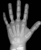
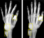
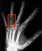
Luan A, von Rabenau L, Serebrakian AT, Crowe CS, Do BH, Eberlin KR, Chang J, Pridgen BC. Machine Learning-Aided Diagnosis Enhances Human Detection of Perilunate Dislocations. Hand (N Y). 2025 Jan 15:15589447241308603. doi: 10.1177/15589447241308603. Epub ahead of print. PMID: 39815415; PMCID: PMC11736725.
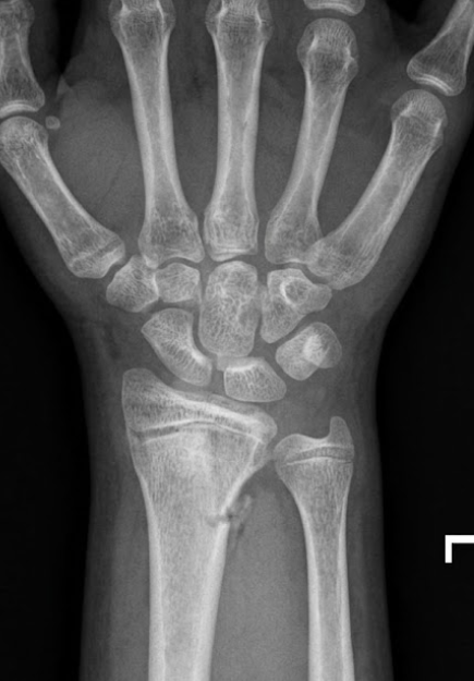
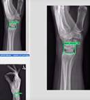
Pridgen B, von Rabenau L, Luan A, Gu AJ, Wang DS, Langlotz C, Chang J, Do B. Automatic Detection of Perilunate and Lunate Dislocations on Wrist Radiographs Using Deep Learning. Plast Reconstr Surg. 2024 Jun 1;153(6):1138e-1141e. doi: 10.1097/PRS.0000000000010928. Epub 2023 Jul 17. PMID: 37467052.
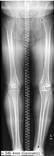
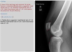
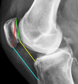
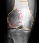
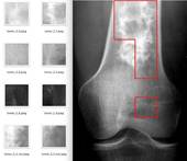
Larson N, Nguyen C, Do B, Kaul A, Larson A, Wang S, Wang E, Bultman E, Stevens K, Pai J, Ha A, Boutin R, Fredericson M, Do L, Fang C. Artificial Intelligence System for Automatic Quantitative Analysis and Radiology Reporting of Leg Length Radiographs. J Digit Imaging. 2022 Dec;35(6):1494-1505. doi: 10.1007/s10278-022-00671-2. Epub 2022 Jul 6. PMID: 35794502; PMCID: PMC9261153.
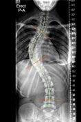
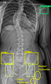
Ha AY, Do BH, Bartret AL, Fang CX, Hsiao A, Lutz AM, Banerjee I, Riley GM, Rubin DL, Stevens KJ, Wang E, Wang S, Beaulieu CF, Hurt B. Automating Scoliosis Measurements in Radiographic Studies with Machine Learning: Comparing Artificial Intelligence and Clinical Reports. J Digit Imaging. 2022 Jun;35(3):524-533. doi: 10.1007/s10278-022-00595-x. Epub 2022 Feb 11. PMID: 35149938; PMCID: PMC9156601.