The NH2-Terminus of beta-catenin contains a potential recognition site for
the serine/threonine kinase glycogen synthase kinase (GSK)-3beta. Mutation
of this site increases the stability of beta-catenin in the adenomatous
polyposis coli (APC) protein complex. DeltaGSK-beta-catenin accumulates in
microtubule-associated APC protein clusters at the tip of cell extensions.
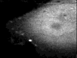
Movie A: The dynamics of these clusters are visualized by time lapse
confocal microscopy of MDCK cells expressing a fusion of
DGSK-beta-catenin with green fluorescent protein (GFP).
56 Frames collected every 40 sec, total time of movie: 37 min
(For a 750 KB QuickTime movie of better quality, click here)
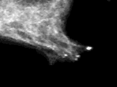
Movie B: Time lapse confocal microscopy after co-transfection of MDCK cells
with GFP-DGSK-beta-catenin and GFP-tubulin. The DGSK-beta-catenin cluster
are localized at the end of microtubule filaments.
94 Frames collected every 27 sec, total time of movie: 42 min.
(For a 2.6 MB QuickTime movie of better quiality, click here)
GFP-fusion constructs made by Eugenio L. de Hostos, Department of Biochemistry and Cell Biology, Rice University. Transfections
and imaging by Angela I. Barth, Department of Molecular Physiology, Stanford University School of Medicine, with the assistance of
Cynthia L Adams, Stephen J. Smith, and W. James Nelson, Stanford.
Laser confocal microscope designed and built by Stephen J. Smith (see Adams, Nelson, and Smith, 1996, J. Cell Biol. 135:
1899-1911).
![]() Additional movies can be found in Dr.
Eugenio de Hostos' Movie Web Page
Additional movies can be found in Dr.
Eugenio de Hostos' Movie Web Page