| Monopartite, positive sense, single stranded RNA.
5' capped, 3' polyadenylated 10-12 kb long Cytoplasmic replication Nonstructural proteins encoded at the 5' end |
Schematics by Steve Folder |
Capsid-
|
Enveloped T=3 icosahedral nucleocapsid 40-70 nm diameter Comprised of multimers of 1 gene product Spherical |
 |
Envelope-
Tightly bound to capsid
2 peplomers: E1 and E2 are responsible for viral specificity
Envelope acquired by budding
Translation & Protein Manipulation-

- Image is public domain
Because RNA is positive sense, it is immediately able to be translated by cellular ribosomes and tRNA.
Two-thirds of the RNA on the 5' side are translated into a polyprotein, which subsequently cleaves itself into four non-structural proteins (NSP).
NSP1- methylation and capping of viral RNA
NSP2- helicase and protease
NSP3- converts RNA replicase to plus strand replicase
NSP4- replicase
Following translation of the non-structural viral proteins, the positive sense genome is transcribed tomproduce a negative sense copy. From this negative sense copy, a full length positive sense genome is produced for packaging, and a sub-genomic 3' mRNA fragment encoding the structiral proteins is made.
The remaining 3' third of viral RNA is translated into another polyprotein that encodes all of the stuctural proteins. Alphavirinae's polyprotein is autolytic for the C protein, which is cleaved while the protein is in the cytosol. The remaining envelope proteins insert into the endoplasmic reticulum, where they are cleaved by host-cell signalase. Following glycosylation the envelope proteins are released back into the cytosol, where they bind to the cellular membrane.
Rubivirinae goes about cleavage in a slightly different manner by initially cleaving the E1 protein from E2 in the cytosol, The E1 and transmembrane proteins are cleaved by host-cell signalase in the endoplasmic reticulum. The E2-Capsid protein is cleaved by host-cell signalase and immediately associates with the membrane for viral assembly. The difference in management and manipulation of the NSP polyprotein, and gene order, in addition to vector trasferability, are the major criteria used to separate the alphavirinae from the rubivirinae.
C- capsid
E1, E2, (E3)- Envelope peplomer
Small transmembrane protein
The nucleocapsid protein spontaneously assembles with inserted envelope proteins to form a tightly-bound nucleocapsid-envelope complex. This complex then selectively packages genomic RNA over the sub-genomic mRNA encoding the NSP polyprotein. This phenomenon has been observed in in vivo studies, but has not been repeated in vitro, suggesting that the mechanism of genome uptake is not fully understood.
The virus acquires its envelope by budding through the cellular membrane. Alphavirinae, which bud through cytoplasmic membranes, are further distinguished from the rubivirinae, which bud through intracytoplasmic membranes.
Infection Cycle

- Image is public domain
The infection cycle of togaviridae begins when the virion is taken up into cellular vessicles by receptor-mediated endocytosis. A pH drop caused by the merging of the carrier vessicle with a cellular lysosome digests nucleocapsid proteins and releases the genomic RNA to the cytosol where replication begins. The positive sense RNA is immediately translated as describes above. New virions spontaneously assemble following viral translation and replication. The virion then buds through cellular membranes.
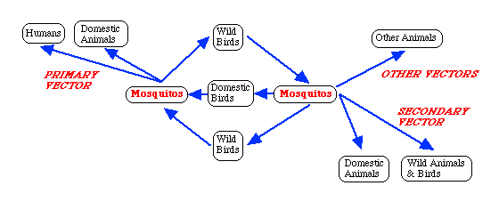
Alphavirinae are considered to be zoonoses of man, as their main host are domesticated and wild animals. Parasitized by mosquitos, the virus is taken up in the animal host's blood. The virus replicates within the mosquito vector before it is introduced into a human host through mosquito bite. The virus is introduced into the capilarries, and replicates in the vasular endothelium before being spread through the blood to other target tissues including muscles, joints, skin, and brain.
Rubivirinae lack animal reservoirs, and are spread between man easily. The the virus is spread via aerosol, and enters the body trough inhalation. It establishes infection in the upper respiratory tract, and spread to the lymph nodes. The virus incubates for ~18 days and manifests itself as a rash with fever.
