| Image | Description | Courtesy of |
|---|---|---|
| Electron micrograph and computer model | Clear representation of the Capsid | Linda Stannard, University of Capetown |
 |
Negative stain TEM at x250000 (About a dozen Adenovirus virions) | Dr. Stewart McNulty, Veterinary Sciences, Queens University (For Educational Purposes Only) |
 |
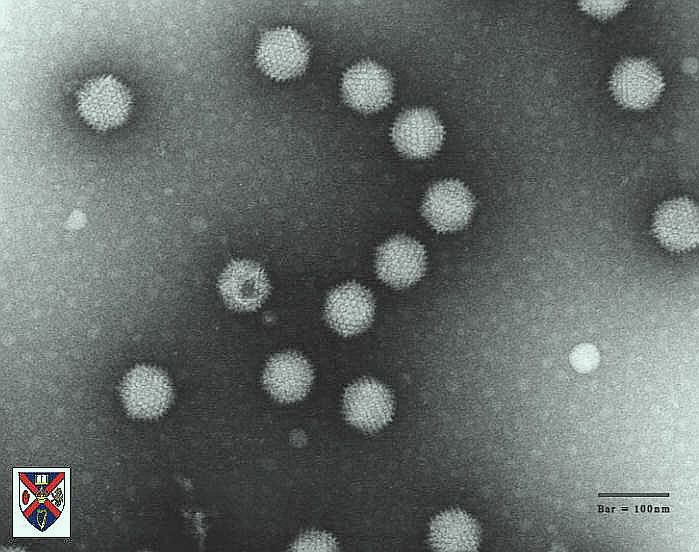
Negative stain TEM, Bar=100nm. Adenoviruses (large, with definitive icosahedral shape) and Parvoviruses (small, more spherical) from the stool sample of an individual with gastroenteritis. | F.P. Williams, MCEARD, and the Evironmental Protection Agency (EPA) |
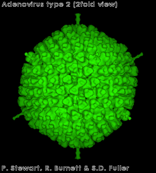 |
Cryo-EM of an idenovirus in the 2-fold access of the icosahedron. | Stewart, P.L., Burnett, R.M. and Fuller, S.D. (1991) Image reconstruction reveals the complex molecular organization of adenovirus. Cell 67:145-167 Stewart, P.L., Fuller, S.D. and Burnett, R.M.(1993) Differnce imaging of adenovirus: bridging the resolution gap between X-ray crystallography and electron microscopy EMBO Journal 12:2589-2599 |
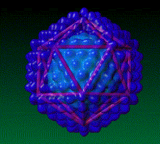 |
Electron microscope reconstruction of adenovirus, showing its icosahedral nature. | © 1992 David S. Goodsell, TSRI |