|
Prediction and Early Detection of Damage Accumulation in Human Tendons |
Investigators: Dennis R. Carter, PhD and Gary S. Beaupré, PhD
Project Staff: Tishya A. Wren, PhD; Scott A. Yerby, PhD; R. Lane Smith, PhD; and Garry Gold, MD
Purpose: Many individuals suffer from tendon injuries, especially injuries of the Achilles tendon, and these injuries are becoming increasingly common as the population ages and becomes more involved in health and fitness activities. We hypothesize that both acute and chronic tendon injuries result from the accumulation of loading-induced damage. This study will examine the injury mechanism of damage accumulation in human tendons with a particular focus on the Achilles tendon.
Objectives: Our long term objective is the prevention of tendon injuries, especially injuries of the Achilles tendon, through an improved understanding of damage accumulation in tendons and through the prediction and early detection of damage accumulation. The specific aims of this study are: (1) to characterize loading-induced damage accumulation in human Achilles tendons in vitro: (2) to develop a computer model that can predict the amount of damage accumulated in a tendon; and (3) to identify medical imaging techniques that can detect early stages of damage accumulation in human Achilles tendons in vivo.
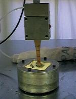
Methodology: This study involves a combination of in vitro testing, computer modeling, and clinical investigation. To characterize damage accumulation in the absence of a healing response, we will perform mechanical testing on human Achilles tendons in vitro. We will incorporate the knowledge gained from the in vitro studies into a previously developed tendon adaptation model to better understand and predict damage accumulation in human tendons. Finally, we will use conventional magnetic resonance imaging, short echo time magnetic resonance imaging, and high frequency ultrasound to image human Achilles tendons in vivo.
Findings: Our models suggest that tensile strains are an important mechanical parameter governing tendon adaptation and injury. Our models demonstrate how tendons may adapt their mechanical properties in response to cyclic strains, thereby reducing high strains that might lead to injury in the absence of this adaptive response. Our models also indicate, however, that there may be physical limits on the extent to which a given tendon can adapt. A tendon that reaches these limits is at increased risk of injury since it will be exposed to higher strains. Our mechanical testing suggests that the Achilles tendon is one such tendon. We have shown that the Achilles tendon has material properties similar to other tendons even though it is exposed to much higher in vivo stresses. The Achilles tendon therefore experiences much higher strains than most other tendons. We believe this helps to explain the high incidence of Achilles tendon injuries. To improve the prevention of loading-induced injuries, we are imaging Achilles tendons in vivo using new MRI techniques. These new techniques allow us to capture much more signal from tendons than is obtained during conventional MRI. This additional signal may allow earlier detection of damage accumulation in tendons and therefore early intervention to prevent injury.
Peer-Reviewed Articles:
Wren TAL and Carter DR (1998) A microstructural model for the tensile constitutive and failure behavior of soft skeletal connective tissues. J. Biomech. Eng. 120, 55-61.
Wren TAL, Beaupré GS, Carter DR (1998) A model for loading-dependent growth, development, and adaptation of tendons and ligaments. J Biomech 31, 107-114.
Wren TAL, Beaupré GS, Carter DR (2000) Tendon and ligament adaptation to exercise and immobilization. Journal of Rehabilitation Research & Development 37, 214-224.
Wren TAL, Beaupré GS, Carter DR (2000) Mechanobiology of tendon adaptation to compressive loading through fibrocartilaginous metaplasia. Journal of Rehabilitation Research & Development 37, 135-151.
Wren TAL, Yerby SA, Beaupré GS, Carter DR (2000) Interpretation of calcaneus dual energy x-ray absorptiometry measurements in the assessment of osteopenia and fracture risk. Journal of Bone and Mineral Research 15, 1573-1578.
Funding Source: VA Rehabilitation Merit Review project