A HISTORY OF THE SKELETON
![]()
"It is very clearly apparent from the admonitions of Galen how great is the usefulness of a knowledge of the bones, since the bones are the foundation of the rest of the parts of the body and all the members rest upon them and are supported, as proceeding from a primary base. Thus if any one is ignorant of the structure of the bones it follows necessarily that he will be ignorant of very many other things along with them."-- Niccolo Massa, 1559
Physicians from antiquity through the Renaissance discussed the form and function of the skeleton, as the hardest part of the body. Beginning with Galen, the investigations of the skeleton followed a certain pattern. Physicians were primarily impressed with the hardness of the bone and saw its necessity for the structural integrity of the body. Galen observed:
"To protect the system completely, it was better for it to consist of many bones, and further, of bones just as hard as they are ... Nature consequently did not merely entrust its defense to the skin, as she did for the parts in the abdomen, but first, before the skin was put on, she invested it with bone like a helmet."
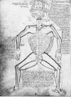
This perspective is fully evident in medieval images of the skeleton that emphasize its ability to shape the body. Look below to see how the skeleton appears in the late Middle Ages.
 Galen
also drew a series of logical conclusions about the shape and weight of specific
bones, observing that the femur was the largest bone in order to sustain the
body's weight, and noting the concavity and convexity of bones that "must
articulate with one another, particularly if the bones are
large." He also argued that it was made from sperm because of
its pale color. As late as 1620, the Scottish physician John Moir could
lecture to his students: "Bone is ... generated out of semen, fat and
earth by the power of heat and the innate spirit." Each succeeding generation after Galen relied
heavily upon his knowledge. In the eleventh century, Avicenna offered a
humoral explanation of bone as primarily made of earth. He based his
conclusion on the fact that bones were cold and dry, like the earth
itself. He qualified this comment with an interesting experiment:
"The bone ... is however moister than hair, because bone is derived from the blood, and its fume is dry, so that it dries up the humors naturally located in the bones. This accounts for the fact that many animals thrive on bones, whereas no animal thrives on hair-- or at least it would be a very exceptional thing if hair ever did provide nourishment. The proof that bone is moister than hair is that when equal weights of bones and hair are distilled in a retort, more water and oil will flow and less "faex" will remain."
Avicenna also offered the practical advice that the best way to gain knowledge of the skeleton was to see it separated from the rest of the body, an idea that became common practice in the Renaissance.
On the whole, however, medieval and early
Renaissance anatomists had less to say about the skeleton than many other parts
of the body. It seemed to them deceptively simple and self-evident in ways
that le ss visible structures
did not. After all, it was not primarily learned physicians who were
interested in the skeleton but surgeons and bone-setters -- less learned
practitioners who dealt directly with the ordinary and extraordinary health
problems associated with broken bones. In the first decades of printing,
many early almanacs and surgical manuals included elaborate diagrams of the
skeleton to assist practitioners and patients in knowledge of the body.
Look at the two images here for an example.
At the end of the fifteenth century, renewed
interest in dissection led to closer inspection of skeletons. Published
anatomies during the Renaissance display
one of the common problems of this era th
 at was especially apparent in discussing the
skeleton -- a complex structure with numerous parts. What was the proper
name for each bone? Jacopo Berengario da Carpi addresses this problem by
including all possible names at the end of the century: Greek, Arabic and
Latin: "This is properly called the hand ... because
from this part almost all handicrafts emanate. Between this and the second
part is a juncture composed of many bones called in Arabic raseta and ascam
and in Greek carpus." Berengario included detailed diagrams in his
popular anatomy that now focused on singular parts of the skeleton, as in this
illustration here.
Berengario's crude woodblocks could not compare to
the hand-drawn illustrations of his
 contemporary, the artist and anatomist Leonardo da
Vinci. Look at Berengario's images of the skeleton above and compare it to
Leonardo's beautiful, highly geometrized drawings of the skull and ribs.
Both dissected, yet they saw the world very differently. Leonardo make
careful notes to himself about the importance of drawing the skeleton from
multiple perspectives: "Make a demonstration of these ribs in which the thorax is shown
from within, and also another which has the thorax raised and which permits
the dorsal spine to be seen from the internal aspect. Cause these 2 scapulae
(spatole) to be seen from above, from below, from the front, from behind,
and forward."
 Confusing terminology was not the only
problem facing Renaissance anatomists; they also discovered that their
descriptions diverged considerably from Galen's, because he had frequently taken
the similarity between human and animal anatomy to be an exact
correspondence. "In the larger hand there are thirty bones,"
stated Alessandro Achillini in 1520. "There would be thirty-one
if the ninth of Galen was included, but that, however, is a monkey bone."
By the time Andreas Vesalius published On the Fabric of the Human Body
(1543), he could cite numerous errors of Galen in the number and shape of the
bones, though he, too, continued to identify many animal parts as belonging to
humans. Leonardo played with the confusion between human and animal
anatomy by drawing a fanciful foot of a beat based on a human one -- an
interesting reversal of the common trend.
There were many things that Renaissance medical practitioners did not fully understand about bones, though Renaissance anatomy theaters were filled with articulated skeletons by the late sixteenth century, such as the one Vesalius prepared in 1546 that can still be seen today at the University of Basel. They knew that bones had differing degrees of density, flexibility and motility. But they had a very limited understanding of more complicated questions such as the relationship between the vertebrae and the spinal cord.
"There are thirty vertebrae. But the round bone upon which the head rests makes thirty-one when it is included in the number of the vertebrae. There are seven vertebrae of the neck; they are slender but have a larger cavity or aperture, however, and are hard and firmly joined to each other."
Alessandro Achillini then found himself wondering in 1520 how it all worked. "Or does the tenth vertebra have two pieces or processes? Or do the processes ascend above and descend below the tenth vertebra? Or does the tenth vertebra have two cavities?" It was much simpler to say, as Nicolo Massa did in 1559, "Nature has made the spine for animals to be like the keel of the body that is necessary for their life; for it is thanks to the spine that we can walk erect and each of the other animals can walk in the posture that is the better one for it."
 Very few Renaissance anatomists, other than
Vesalius, paid such close attention to the skeleton as a whole, preferring to
pay special attention to parts such as the skull, that was an object of great
fascination because of the continued interest in physiognomy. In the
majority of instances, physician's best insights were closely connected to their
interests in other parts of the body. For example, it is hardly surprising
that William Harvey in his 1653 anatomy should pay special attention to the
sternum, given his detailed exploration of the heart and lungs. He wrote:
"There are three uses for the sternum: rampart for the heart and
vitals, binding for the ribs, support for the membranes of the mediastinum.
Sometimes it protrudes outward, out cheated, origin of gibbosity. The sternum
is made up of 6 or 7 bones, more in children and fewer occur in old age."
Even though many medical practitioners did not puzzle over the skeleton to the same degree that they did with other parts of the body, they all recognized their cultural as well as scientific importance. By the end of the sixteenth century, skeletons had become the quintessential image of anatomy. But they also continued to be an image of death, in the form of a grim reaper brought to life by the skill of the anatomist.
QUESTIONS: WHY DID KNOWLEDGE OF THE SKELETON REMAIN RELATIVELY STABLE? HOW HAS THE SKELETON DEFINED OUR HUMANITY IN UNIQUE WAYS?